
To Schedule An Appointment
Call 405-310-6977
Shon Cook, M.D.
Certified by the American Board of Neurological Surgeons
Specializing in Minimally Invasive Neurosurgery

Important Notice: Our office is now in its new location at 10001 S. Western Ave, Suite 101 in Oklahoma City. The Contact and Map page has been updated. We are only in the office on Mondays, but you can drop off images and records any day of the week and we will get them on Monday.
Dr. Cook's Brain Surgery Patients
Right after surgery, Dr. Cook's brain surgery patients look like they have not had surgery.


Both of these patients had brain surgery by Dr. Cook about 2 weeks before these pictures were taken. Typically, patients go home the day after surgery, looking like they have not had surgery.
Dr. Cook's Incisions
Below are examples after surgery using the keyhole approaches Dr. Cook uses for tumors, aneurysms, trigeminal neuralgia, shunts, endoscopy, and Chiari malformation surgeries

Dr. Cook’s VP Shunt Incision
(hair down)

Dr. Cook’s VP Shunt Incision
(hair pulled back, glue still on)
Comparisons
Below are examples of people’s incisions after the traditional open craniotomy for the same pathology. These images are publicly available on the internet. Clicking the picture will take you to the posting page.*

VP Shunt Incision
from internet

VP Shunt Incision
from internet

Dr. Cook’s Aneurysm Incision
(hair down)

Dr. Cook’s Aneurysm Incision
(hair pulled back, glue still on)

Aneurysm Incision
from internet

Aneurysm Incision
from internet

Dr. Cook’s Left Eyebrow Incision
(1 week)
.jpg)
Left Frontal Incision
from internet

Dr. Cook’s Right Forehead Incision
(3 weeks)
Right Frontal Incision
from internet

Dr. Cook’s Retrosigmoid Incision
(surgery day with glue on)

Retrosigmoid Incision
from internet

Dr. Cook’s Chiari Incision
(surgery day with glue on)
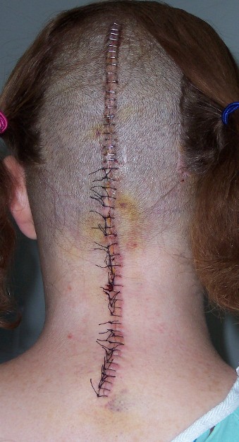
Chiari Incision
from internet
*All of the comparison images used on this website are found in various places throughout the internet and are believed to be within the public domain. Images used are believed to be within our rights as stated within the US Copyright Fair Use Act. If you think any content on this website infringes your copyright or violates your privacy please let us know, and we will remove the images in question. You can send correspondence to shoncookmd@gmail.com